May 22, 2017
An international team of researchers, affiliated with UNIST has developed a new type of adaptive-optics (AO) technology for brain research.
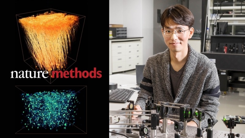
This study has been jointly conducted by Professor Jung-Hoon Park of Life Sciences at UNIST in collaboration with Professor Meng Cui of Electrical and Computer Engineering at Purdue University’s West Lafayette, Indiana, United States.
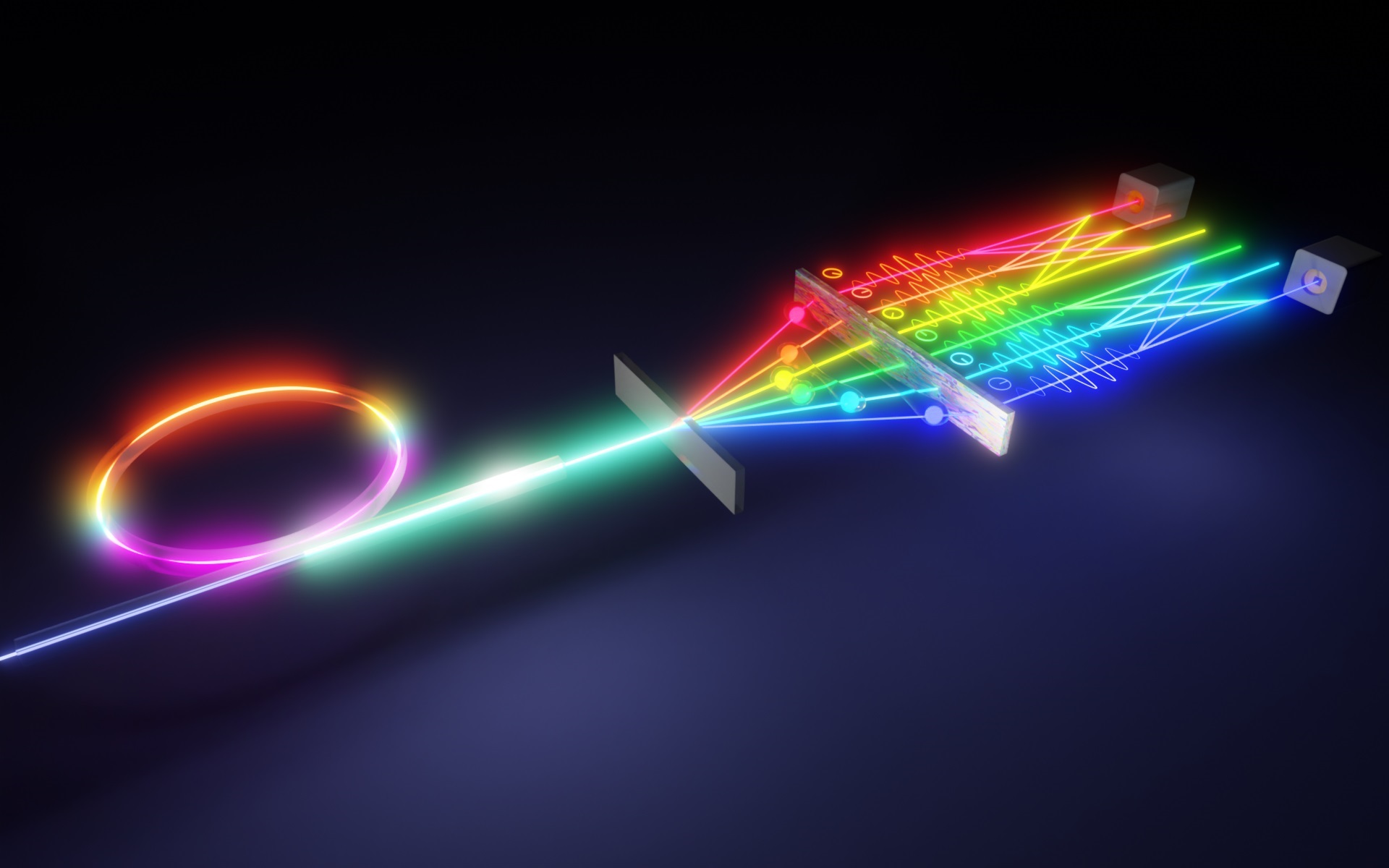
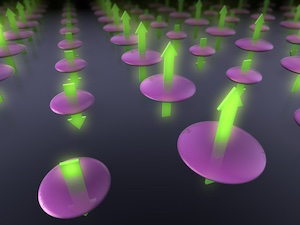
In the study, the research team has developed Multi-Pupil Adaptive Optics (MPAO) to perform high-resolution time-lapse imaging to study changes in functioning brain cells. According to the research team, the idea here is that studying subtly changing images can provide new insights into how the brain works.
This newly-developed AO system enbables simutaneous wavefront correction over a large imaging field-of-view (FOV). In the current implementation, MPAO improves correction area by approximately an order of magnitude over that of conventional methods and allows up to 32 MHz voxel rate at the limit of the laser scanning hardware.
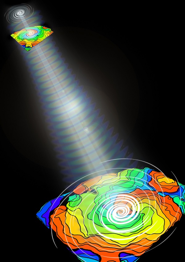
The capability of independent wavefront control for MPAO also enables high-speed nonplanar imaging of 3D dynamics. In the study, the research team applied MPAO to in vivo dynamic imaging of microglia and blood vasculature, and structural and calcium imaging of neuronal network in mammalian brain.
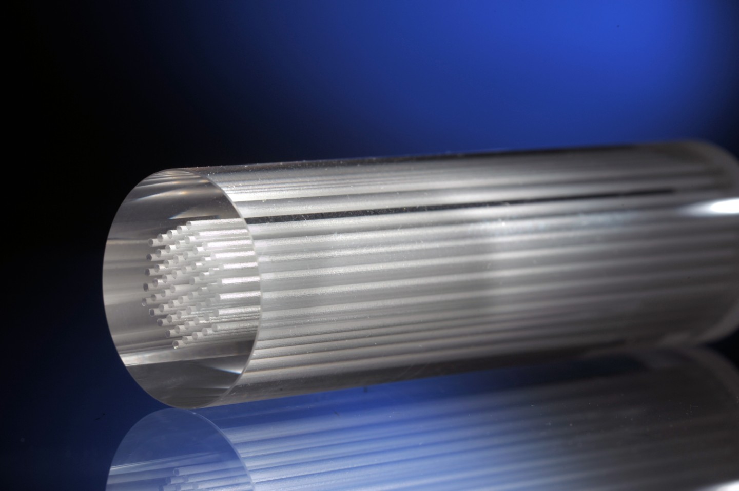
The prototype multi-pupil AO system includes 9 deformable mirrors and a prism array containing 9 faceted segments. Each segment produces its own image corresponding to a different part of a microscope’s field of view. The approach improves the area of AO correction by 9 over a single-deformable-mirror approach; the field of view of the new system is 450 × 450 μm2. The system images voxels at rates up to 32 MHz, limited by the laser-scanning hardware.
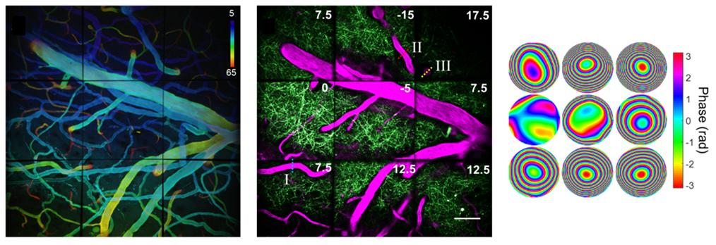
The system images voxels at rates up to 32 MHz, limited by the laser-scanning hardware.
“To better understand brain activity, we must look directly at the dynamic connection between brain cells distributed over a wide area,” says Professor Park. “High-resolution in-vivo imaging of dendritic spines is also of great importance in neuroscience because you want to measure these numerous neurons simultaneously at very high speed and also at high spatial resolution.”
The results of the study have been published in the May issue of the renowned scientific journal, Nature Methods (IF=25.328). It was carried out with the support of the National Institutes of Health (NIH) and Purdue University.
Journal Reference
Jung-Hoon Park et al., “Large-field-of-view imaging by multi-pupil adaptive optics
Jung-Hoon Park,” Nature Methods, (2017).